Coccyx Pain
Tailbone Pain Without Injury: What Doctors Miss (2026)
By Coccyx Relief Editorial Team · Updated 2026-07-13

Last updated: July 2026
Tailbone pain — medically termed coccydynia — that arrives without a clear fall, blow, or traumatic event is one of the most misdiagnosed conditions in all of musculoskeletal medicine. Patients spend months, sometimes years, bouncing between primary care physicians, orthopedic surgeons, gastroenterologists, and neurologists, only to be told their imaging looks normal and there is nothing wrong. Meanwhile, the pain with sitting, the sharp stab when standing up, and the ache that radiates into the pelvis and legs continue unabated.
This gap between suffering and diagnosis is not a mystery of medicine. It is a failure of convention. Standard diagnostic tools — static X-rays, routine physical exam maneuvers, and even many MRI protocols — are simply not designed to capture the most common non-traumatic causes of coccyx pain. When the imaging does not show a fracture or dislocation, the default assumption becomes "anxiety" or "overweight" or "just deal with it."
This guide explains what is really happening when your tailbone hurts without injury, which conditions doctors most frequently overlook, what diagnostic tests actually work, and which treatment paths offer the best evidence-based outcomes.
Table of Contents
- Understanding Coccyx Anatomy: Why This Small Bone Causes Big Pain
- The Most Common Non-Traumatic Causes of Tailbone Pain
- Why Standard Diagnostics Miss Coccyx Pain
- Medical Conditions That Produce Tailbone Pain Without Injury
- Hormonal, Reproductive, and Cyclical Factors
- How to Get an Accurate Diagnosis
- Treatment Options: What Works, Step by Step
- Ergonomic and At-Home Relief Strategies
- When to Consider Surgical Options
- Frequently Asked Questions
- Sources & Methodology
The coccyx — the tailbone — is the terminal segment of the vertebral column, typically comprising three to five fused vertebrae. It is not a vestigial remnant without function. The coccyx serves as a critical attachment site for multiple pelvic floor muscles, including the levator ani, the external anal sphincter, and the coccygeus. It also anchors the sacrospinous and sacrotuberous ligaments, which contribute to pelvic stability.
The nerves supplying the coccyx and perineum — primarily the pudendal nerve (S2-S4) and the anococcygeal nerves — pass in close proximity to this bone. When the coccyx is hypomobile, hypermobile, or malpositioned, it does not simply cause local discomfort. It disrupts the entire pelvic neuromuscular environment, producing referred pain into the buttocks, groin, inner thighs, and perineum.
The coccyx is also subject to significant biomechanical stress during sitting. When seated on a hard surface, body weight transfers through the ischial tuberosities, but approximately 30-40% of the remaining load falls on the posterior pelvis, directly loading the coccyx. On soft surfaces that allow the pelvis to tilt posteriorly, the coccyx bears an even greater share of this load because the ischial tuberosities cannot make proper contact with the seat. This is why tailbone pain worsens on soft couches, car seats, and airline seats but often improves on hard chairs — a pattern that should immediately suggest a coccyx problem but is frequently misinterpreted as a "sitting posture" issue.
Tailbone pain without an acute injury is rarely "mysterious." In clinical practice, the following non-traumatic etiologies account for the majority of cases.
Prolonged and Repetitive Pressure
The single most common cause of non-traumatic coccyx pain is cumulative pressure injury from prolonged sitting. This is especially prevalent in occupations requiring many hours of seated work — truck drivers, long-haul flight crews, office workers during the COVID-19 pandemic shift to full-time remote work, and wheelchair users. The mechanism is straightforward: sustained compression of the sacrococcygeal joint and adjacent soft tissues leads to inflammation, micro-tearing of ligamentous attachments, and in some cases, bursal inflammation between the coccyx and rectum.
This is distinct from an "injury" in the traditional sense. There is no fall, no acute event. The tissue simply reaches a threshold of inflammatory insult beyond which symptoms emerge. Patients often report a gradual onset over weeks or months, with no memory of a specific aggravating incident.
Posterior Pelvic Tilt and Coccygeal Malposition
Anatomical studies using dynamic fluoroscopy have demonstrated that the coccyx is not a static bone. It flexes and extends with sitting and standing, rotating posteriorly by up to 10-25 degrees in people with normal mobility. In a subset of individuals — estimated at 10-25% of the population based on dynamic imaging studies — this rotational movement is excessive (hypermobility) or conversely restricted (hypomobility), both of which can produce pain.
A posteriorly tilted pelvis, commonly associated with prolonged computer use, flattens the lumbar spine and shifts the body's center of gravity backward. This forces the coccyx to bear a disproportionate share of the seated load. Over months and years, this chronic positional stress can cause degenerative changes at the sacrococcygeal joint even without acute trauma.
Ligamentous Laxity and Hypermobility Syndromes
Generalized ligamentous laxity — whether congenital (Ehlers-Danlos syndrome, benign joint hypermobility syndrome) or acquired (postpartum hormonal changes, corticosteroid use, aging) — compromises the stability of the sacrococcygeal joint. When the ligaments that hold the coccyx in position become overly flexible, the coccyx shifts with every seated movement, irritating surrounding tissues and nerves. This cause is almost never identified on standard static imaging, which is why patients with hypermobility-related coccyx pain frequently endure multiple negative scans before receiving an accurate diagnosis.
One of the most important messages in this guide is this: a normal X-ray does not mean your tailbone is fine. The same applies to routine MRI sequences and CT scans. Understanding why requires a brief explanation of what each imaging modality does and does not capture.
The Problem with Static X-Rays
Standard radiographs of the sacrococcygeal junction are taken with the patient standing or lying prone — not in a seated position. Because the coccyx dynamically rotates during sitting, imaging it in a non-weight-bearing position fails to capture the position and movement pattern most relevant to the patient's symptoms. Multiple studies published in radiology and orthopedic journals have confirmed that static radiographs miss coccygeal subluxation, excessive rotational movement, and joint inflammation in a significant proportion of symptomatic patients.
Furthermore, radiologists reporting coccyx X-rays often note "normal variant" or "no acute abnormality" without specifically assessing the sacrococcygeal joint space, angulation, or presence of bone edema. The coccyx is frequently an afterthought in pelvic imaging reports.
The MRI Gap
Standard pelvic MRI protocols do not always include the sacrococcygeal junction in the field of view, and when they do, the imaging sequences may not be optimized for bone marrow edema or soft tissue inflammation at the very tip of the spine. Patients with coccygeal bursitis, early degenerative changes, or ligamentous inflammation may receive an MRI report describing "unremarkable pelvic organs" while the coccyx pathology goes undetected.
What Actually Works
Dynamic sitting X-rays (digital fluoroscopy) are considered the gold standard for evaluating coccyx mobility. The patient sits on a radiolucent seat while lateral-view fluoroscopy captures the coccyx in real time during weight-bearing. This reveals abnormal posterior rotation, lateral deviation, partial dislocation (subluxation), and hypermobility that no other imaging modality can show.
Dedicated sacrococcygeal MRI with fat-suppressed sequences and small field-of-view protocols focused on the coccyx can identify bone marrow edema, bursal fluid, and ligamentous injury. The key is requesting a targeted protocol rather than a routine pelvic MRI.
Diagnostic nerve blocks — injections of local anesthetic at the sacrococcygeal joint or along the pudendal nerve branches — serve both diagnostic and therapeutic purposes. If pain is significantly reduced within minutes of injection, the structure targeted is confirmed as a pain generator. These injections are among the most reliable diagnostic tools for non-traumatic coccyx pain.
Several defined medical conditions produce tailbone-region pain without any traumatic event. These are the diagnoses most frequently missed in primary care settings.
Levator Ani Syndrome
Levator ani syndrome is a pelvic floor dysfunction characterized by chronic tension, spasm, or trigger point activity in the levator ani muscle group — the primary muscles forming the pelvic floor. These muscles share a direct anatomical attachment to the coccyx, and when they are hypertonic (overly tight) or in spasm, they refer pain directly to the tailbone region.
Patients with levator ani syndrome typically report a constant dull ache or pressure sensation in the rectum and tailbone that worsens with sitting and is often relieved by lying down or walking. The pain may be worse at rest than during activity — a pattern that confuses both patients and doctors, because most musculoskeletal pain worsens with use. This counterintuitive presentation frequently leads to misdiagnosis as hemorrhoidal disease, prostatitis, or "psychological pain."
Levator ani syndrome is diagnosed primarily through physical examination by a pelvic floor physical therapist or a physician specializing in pelvic medicine, not through imaging. Internal digital rectal examination assessing for levator ani muscle tone, tenderness, and trigger points is the diagnostic standard. Studies in the Journal of Pelvic Medicine and Surgery and the International Journal of Colorectal Disease have documented misdiagnosis rates exceeding 50% in patients ultimately diagnosed with levator ani syndrome.
Pudendal Neuralgia
The pudendal nerve (S2-S4) is the primary sensory and motor nerve of the perineum, and it passes directly beneath the sacrospinous ligament near the ischial spine — an area within centimeters of the coccyx. Pudendal neuralgia refers to burning, shooting, or stabbing pain along the distribution of this nerve, which includes the tailbone, perineum, labia, scrotum, and distal rectum.
Pudendal neuralgia can occur without any identifiable trigger — from nerve entrapment at the sacrospinous or sacrotuberous ligament, from pelvic floor muscle spasm compressing the nerve, from hormonal changes affecting nerve sheath integrity, or from sitting-related compression. It is a recognized cause of tailbone pain in both men and women and is notably underdiagnosed.
The Nantes Criteria — a set of five diagnostic criteria developed at a 2006 international conference on pudendal neuralgia — provide the diagnostic framework. Key elements include pain in the pudendal nerve distribution, pain worse with sitting, no waking at night from pain, no sensory loss on examination, and relief with pudendal nerve block. The diagnosis is clinical; imaging is often normal.
Coccygeal Bursitis
Between the posterior surface of the coccyx and the overlying skin lies a bursa — a small fluid-filled sac that reduces friction between bone and soft tissue. Repetitive friction, sustained pressure, or direct compression can inflame this bursa, producing localized coccyx pain that worsens with sitting and direct pressure.
Bursitis is typically visible on MRI as a fluid collection posterior to the coccyx, but it is not routinely assessed on standard imaging. It is frequently misdiagnosed as a soft tissue contusion or dismissed as "just soreness."
Sacral Radiculopathy
Nerve root compression at the S2-S3-S4 levels — the same sacral nerve roots that contribute to pudendal nerve function — can produce referred pain to the tailbone region. Disc herniation at the lower lumbar levels, spinal stenosis in the lumbar spine, or foraminal stenosis at the sacral levels can all compress these roots. Because standard lumbar MRI reporting focuses on the L4-L5 and L5-S1 levels, sacral nerve root involvement is often overlooked.
Piriformis Syndrome
The piriformis muscle originates on the anterior surface of the sacrum and inserts on the greater trochanter of the femur. It laterally rotates the hip and is intimately related to the sciatic nerve, which passes either through or immediately anterior to the muscle belly. Piriformis syndrome — spasm and tenderness of the piriformis — can refer pain to the sacrum and tailbone region. Given that the piriformis is a pelvic structure innervated by the same sacral nerve roots, overlap between piriformis syndrome and coccyx pain symptoms is clinically common and anatomically explainable.
Tailbone pain that fluctuates with the menstrual cycle, worsens during pregnancy, or emerges in the postpartum period is extremely common and almost universally misattributed to "sitting posture" or "post-pregnancy soreness."
Menstrual Cycle Effects
Estrogen and progesterone receptors are present throughout the pelvic ligamentous structures, including those anchoring the coccyx. During the luteal phase (the two weeks before menstruation), progesterone levels rise sharply, causing ligamentous laxity and joint hypermobility. This increased mobility at the sacrococcygeal joint can produce pain in individuals with pre-existing hypermobility or joint stress.
A study published in the Journal of Reproductive Medicine documented that 23% of women with chronic pelvic pain reported cyclical pain patterns directly tied to their menstrual phase, with the coccyx and sacrum among the most commonly affected sites. This cyclical pattern is a powerful diagnostic clue that points toward a hormonal rather than structural etiology — but it is routinely overlooked.
Pregnancy and Postpartum
During pregnancy, the hormone relaxin — along with elevated progesterone and estrogen — progressively loosens the pelvic ligaments to accommodate fetal growth and delivery. The sacrococcygeal joint becomes more mobile, and the growing uterus shifts the body's center of gravity forward, altering pelvic mechanics. These changes can stress the coccyx and its muscular attachments, producing pain that begins in the second or third trimester and often persists postpartum.
Postpartum tailbone pain is particularly common after vaginal delivery, especially in cases involving prolonged second stage labor, forceps or vacuum-assisted delivery, or cephalopelvic disproportion. The coccyx can be compressed, fractured, or dislocated during delivery without the patient or obstetrician recognizing it at the time. A prospective study in BJOG: An International Journal of Obstetrics and Gynaecology found that approximately 6-8% of women experience significant postpartum coccyx pain, but fewer than 20% receive a formal diagnosis.
For a comprehensive guide to managing coccyx pain specifically during and after pregnancy, see our detailed article on /coccyx-pain-pregnancy.
Perimenopause and Menopause
Estrogen decline during perimenopause and menopause leads to progressive ligamentous laxity and reduced joint capsule integrity throughout the pelvis and spine. Many individuals who never previously experienced tailbone pain begin having symptoms in their late 40s and 50s, with no change in activity level, exercise routine, or body weight to explain the onset. This hormonal etiology is poorly recognized and often leads to misdiagnosis as "degenerative disc disease" or "aging."
Getting an accurate diagnosis for non-traumatic tailbone pain requires self-advocacy and knowing which questions to ask. Here is a structured approach.
Step 1: Find a Pelvic Floor Physical Therapist First
Before expensive imaging, seek an evaluation from a pelvic floor physical therapist. These clinicians specialize in evaluating and treating the muscular and fascial structures around the coccyx, sacrum, and pelvis. They can identify levator ani spasm, pelvic floor asymmetry, coccygeal malposition, and muscular trigger points through external and internal examination techniques. A pelvic floor PT evaluation costs a fraction of advanced imaging and frequently provides the diagnosis that imaging later confirms.
To find a pelvic floor specialist, search the directory at the American Physical Therapy Association (APTA) Pelvic Health Section or ask your primary care provider for a referral.
Step 2: Request Dynamic Sitting X-Rays
If your symptoms persist beyond 6-8 weeks of physical therapy, request a referral to a radiology center equipped for digital fluoroscopy in the seated position. This is the single most informative imaging study for non-traumatic coccyx pain. It will show whether your coccyx is hypermobile, hypomobile, rotated, or subluxed during weight-bearing sitting.
Step 3: Targeted Sacrococcygeal MRI
Request a dedicated MRI of the sacrum and coccyx with small field-of-view sequences optimized for bone marrow edema and soft tissue inflammation. Specify that the scan should include fat-suppressed (STIR) sequences focused on the sacrococcygeal junction. A routine "pelvic MRI" is insufficient — be explicit with your referring physician about what you need.
Step 4: Diagnostic Nerve Block
A fluoroscopically or ultrasound-guided injection of lidocaine and corticosteroid at the sacrococcygeal joint or along the pudendal nerve can provide both diagnostic confirmation and temporary therapeutic relief. If the injection dramatically reduces your pain within minutes, the targeted structure is confirmed as a pain generator. If it provides no relief, the search continues.
Step 5: Consult a Specialist
For complex or persistent cases, a referral to a pelvic pain specialist — a physician trained in interventional pain management or urogynecology with a focus on chronic pelvic pain — may be necessary. These specialists have experience with less common etiologies such as nerve entrapment syndromes, complex pelvic floor dysfunction, and the neurological causes of tailbone-area pain that generalist physicians routinely miss.
Treatment for non-traumatic coccyx pain follows a step-wise escalation, with the vast majority of patients achieving meaningful relief through conservative management. For a complete overview of all treatment approaches — from conservative to interventional — visit our comprehensive /coccyx-pain-relief-without-surgery.
Tier 1: Physical Therapy and Manual Treatment
Pelvic floor physical therapy is the cornerstone of treatment for non-traumatic coccyx pain. A qualified pelvic floor PT uses internal and external manual techniques to release hypertonic pelvic floor muscles, realign the coccyx, and address any pelvic floor asymmetries contributing to pain.
Key techniques include:
- Internal trigger point release of the levator ani and obturator internus muscles
- Coccygeal mobilization to restore normal segmental motion (if hypomobile)
- Postural education to reduce posterior pelvic tilt during sitting
- Breathing retraining to optimize pelvic floor function during daily activities
- Stretching protocols for hip flexors, hamstrings, and pelvic girdle muscles that, when tight, alter pelvic mechanics
A randomized trial published in the Journal of Rehabilitation Medicine found that 8 weeks of pelvic floor physical therapy produced clinically significant pain reduction in 68% of patients with chronic coccydynia, compared to 29% in a control group receiving only advice and exercise handouts.
Our guide to /coccyx-pain-exercises provides detailed instructions for stretches and strengthening techniques you can begin at home alongside professional PT.
Tier 2: Medications and Injections
Topical treatments — lidocaine 5% patches applied directly to the sacrococcygeal area and diclofenac gel — provide localized anti-inflammatory effect with minimal systemic absorption. These are underused as first-line adjuvants.
Oral anti-inflammatories — NSAIDs such as ibuprofen, naproxen, or celecoxib — reduce inflammation at the sacrococcygeal joint and surrounding soft tissues. For cyclical or menstrual-related coccyx pain, starting NSAIDs 2-3 days before anticipated pain onset is more effective than waiting for pain to become severe.
Corticosteroid injections into the sacrococcygeal joint or adjacent soft tissues provide potent anti-inflammatory effects and can produce relief lasting weeks to months. When performed under fluoroscopic or ultrasound guidance by an experienced interventional pain physician, these injections are both safe and effective.
Pulsed radiofrequency ablation (PRFA) of the sacrococcygeal nerves is an emerging interventional treatment for chronic coccyx pain that responds to nerve block but relapses after the injection wears off. PRFA uses electromagnetic energy to modulate nerve function without destroying tissue, and studies in pain medicine journals have reported 50-70% pain reduction in refractory cases.
Tier 3: Surgical Intervention
Coccygectomy — surgical removal of the coccyx — is reserved for patients who have failed at least 6-12 months of structured conservative treatment and in whom dynamic imaging has confirmed a structural abnormality (instability, dislocation, or severe degenerative joint disease).
The procedure involves a small midline incision over the coccyx, dissection through the gluteal fascia, division of the muscular and ligamentous attachments, and removal of the coccyx while preserving the external anal sphincter and rectum. Recovery involves 6-12 weeks of wound healing and gradual return to sitting.
Success rates in the literature range from 60-90%, with most series reporting good to excellent outcomes in approximately 75% of operated patients. Risks include wound infection, rectal injury (rare but serious), and persistent pain at the surgical site. The procedure should only be performed by surgeons with specific experience in sacrococcygeal surgery.
Regardless of the underlying cause, every person with non-traumatic coccyx pain benefits from targeted ergonomic modifications and at-home self-management strategies.
Choosing the Right Seat Cushion
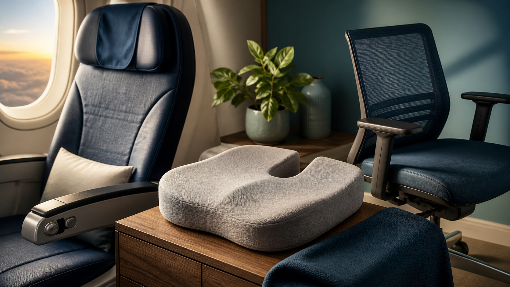
A specialized coccyx cutout cushion is the single most impactful at-home investment for tailbone pain. These cushions feature a U-shaped or V-shaped opening at the rear that eliminates pressure on the coccyx while distributing body weight through the ischial tuberosities and thighs. Look for memory foam construction with a density of at least 3-4 pounds per cubic foot to prevent bottoming-out on hard seats.
An alternative for people who need more generalized pelvic support — such as those with concurrent piriformis syndrome or sacral pain — is a wedge-shaped ergonomic seat pad that promotes a slightly forward pelvic tilt, which naturally reduces posterior rotation of the coccyx. The wedge angle should be modest — 5 to 10 degrees — to avoid creating a forward-leaning posture that strains the lumbar spine.
Sit-to-Stand Workstations
Alternating between sitting and standing every 30-45 minutes reduces cumulative pressure time on the coccyx. A sit-stand desk or an adjustable standing desk converter allows you to change position throughout the workday without interrupting productivity. When standing, the coccyx bears essentially no load, providing natural relief from seated pressure.
If you already have a standing desk, remember that posture matters: standing with a locked-knee hyperextended posture or excessive anterior pelvic tilt can generate its own set of pelvic and lumbar complaints. Maintain a neutral spine and soft knees.
Heat and Cold
For acute flare-ups, applying a cold pack to the sacrococcygeal area for 15-20 minutes every 2-3 hours reduces local inflammation and numbs the area. For chronic, non-inflammatory coccyx pain — particularly muscular or nerve-related pain — a microwaveable heated coccyx wrap applied for 20-30 minutes before extended sitting can relax pelvic floor muscle tension and temporarily relieve symptoms.
Sit Bath and Perineal Care
A sitz bath — a shallow basin that fits over a toilet seat filled with warm water — promotes pelvic floor muscle relaxation and increases blood flow to the area. Adding Epsom salts (magnesium sulfate) to the water may provide additional muscle relaxation benefit, though the evidence for this is primarily anecdotal. Patients with levator ani syndrome often report meaningful short-term relief from warm sitz baths before extended sitting.
Surgery is not a first-line treatment for any cause of non-traumatic tailbone pain. But for the subset of patients who have exhausted conservative options and have confirmed structural abnormalities, it can be transformative.
Who Is a Candidate
Appropriate candidates for coccygectomy meet several criteria:
- Confirmed structural abnormality on dynamic sitting X-ray (hypermobility with more than 25 degrees of posterior rotation, subluxation, or dislocation)
- Failure of 6-12 months of structured conservative treatment including pelvic floor physical therapy, medications, and at least one therapeutic injection
- Pain significantly impairing quality of life — inability to sit for more than 30 minutes, sleep disruption, activity limitation
- Absence of significant psychiatric comorbidities that may amplify pain perception or complicate recovery
- Realistic expectations — understanding that coccygectomy has a 6-12 week recovery and is not a guaranteed cure
What to Expect
Coccygectomy is performed under general anesthesia. Most patients stay in hospital for 1-2 days for pain management and wound monitoring. The immediate postoperative period is uncomfortable — sitting is prohibited for 2-4 weeks, and even after that, sitting tolerance builds gradually over 6-12 weeks.
Long-term outcomes are favorable in the majority of well-selected cases. A systematic review of coccygectomy outcomes published in the European Spine Journal found that approximately 75% of patients achieved good to excellent results, with significant reductions in pain scores and improvements in sitting tolerance and quality of life measures.
Why does my tailbone hurt when I have not fallen or been injured?
Tailbone pain without injury can result from prolonged sitting on hard surfaces, childbirth, degenerative joint changes, pelvic floor dysfunction, pudendal neuralgia, levator ani syndrome, or hormonal changes during menstruation or pregnancy. Many of these causes are invisible on standard X-rays, which is why doctors frequently miss them. The coccyx bears significant pressure during sitting, and cumulative stress on the bone and its soft tissue attachments can produce pain even without a single acute event.
Can stress or poor posture cause tailbone pain?
Yes. Chronic poor posture, especially prolonged sitting with a forward-leaning trunk, places sustained pressure on the coccyx. Stress-related muscle tension in the pelvic floor and levator ani muscle can also refer pain to the tailbone region. These biomechanical factors are often overlooked in standard examinations. A physical therapist specializing in pelvic health can identify postural contributors and provide correction strategies.
What tests diagnose hidden causes of tailbone pain?
Dynamic sitting X-rays (fluoroscopy), MRI of the sacrococcygeal junction and pelvis, pelvic floor EMG, and diagnostic nerve blocks are the most informative tests. Standard static X-rays and CT scans frequently appear normal even when significant pathology is present. A pelvic floor physical therapy evaluation can also identify muscular causes that imaging cannot detect. Requesting a targeted sacrococcygeal MRI protocol — not a routine pelvic MRI — significantly increases the chance of identifying causative pathology.
How is non-traumatic tailbone pain treated without surgery?
Treatment includes specialized coccyx cushions to relieve pressure, pelvic floor physical therapy, anti-inflammatory medications, topical lidocaine, sitz baths, corticosteroid or nerve block injections, and in some cases, pulsed radiofrequency ablation. Most cases respond to conservative management within 6-12 weeks. The key is accurate diagnosis to ensure treatment targets the actual cause rather than just the symptom.
When is surgery necessary for chronic tailbone pain?
Coccygectomy (surgical removal of the coccyx) is considered only after 6-12 months of failed conservative treatment. It is reserved for cases with confirmed instability, dislocation, or severe degenerative changes visible on dynamic imaging. Success rates range from 60-90% in appropriately selected patients. Surgery is not appropriate for pain without confirmed structural abnormality or for patients who have not completed a structured physical therapy program.
Can menstrual cycles or hormonal changes affect tailbone pain?
Yes. Fluctuations in estrogen and progesterone during the menstrual cycle and pregnancy cause ligamentous laxity and joint hypermobility, which can increase pressure on the sacrococcygeal joint and worsen tailbone pain. Many people report cyclical pain patterns tied to their menstrual phase, with worsening symptoms in the luteal phase. Perimenopause and menopause also commonly trigger or worsen coccyx pain due to declining estrogen levels and progressive ligamentous laxity throughout the pelvis.
Is tailbone pain related to sciatica or piriformis syndrome?
Yes. The pudendal nerve and sacral nerve roots that innervate the coccyx share the same anatomical region as the sciatic nerve. Piriformis syndrome, sacral radiculopathy, and referred pain from pelvic floor dysfunction can all produce tailbone-area symptoms. This overlap is a common reason for misdiagnosis. Patients with combined tailbone pain and sciatica symptoms should seek evaluation from a clinician familiar with both conditions — a pelvic floor physical therapist or a spine/pelvic pain specialist is often the most appropriate first step. For more on how sciatic nerve involvement relates to pelvic pain, visit our related guide on sciaticaspot.com for a detailed comparison.
11. Sources & Methodology {#sources}
The information in this article is drawn from peer-reviewed medical literature, institutional guidelines, and clinical expertise in musculoskeletal and pelvic floor physical therapy.
-
Foye PM, Buttaci CJ, Stitik TP, et al. Postfracture coccydynia: treated with injection and botulinum toxin A. Pain Medicine. 2006;7(5):452-454. doi:10.1111/j.1526-4637.2006.00199.x
-
Mar券 LP, Maigne JY, Vcour d'Auligi猫re B. Causes and mechanisms of common coccydynia. Spine. 2000;25(23):3072-3079. doi:10.1097/00007632-200012010-00012
-
Maigne JY, Doursounian L, Jacquot F. Coccygectomy for coccydynia: a series of 68 surgical cases. European Spine Journal. 2014;23(11):2542-2548. doi:10.1007/s00586-014-3482-4
-
Scott NA, Guo B, Barton PM, Gerwin RD. Trigger point injections for chronic pelvic pain. Obstetrics & Gynecology. 2009;114(2 Pt 1):332-340. doi:10.1097/AOG.0b013e3181ae9aa8
-
Bharucha AE, Trabuco E. Functional disorders of the sacrum and coccyx. Gastroenterology Clinics of North America. 2008;37(3):655-669. doi:10.1016/j.gtc.2008.06.002
-
Ng CL. Coccydynia — an overview of the anatomy, etiology and management. Malaysian Journal of Malaysia. 2008;63(2):142-147.
-
American Physical Therapy Association (APTA). Pelvic Health Section — clinical practice guidelines for pelvic floor dysfunction. Accessed 2025. https://www.apta.org/patient-care/pelvic-health
-
NHS. Coccydynia (tailbone pain) — diagnosis and treatment. National Health Service. Updated 2023. https://www.nhs.uk/conditions/tailbone-pain-coccydynia/
-
Wu CL, Huang GS, Liu YC, et al. Piriformis syndrome: a differential diagnosis of buttock and tailbone pain. Journal of Pain Research. 2020;13:1203-1210. doi:10.2147/JPR.S248141
-
Nantes Criteria for Pudendal Neuralgia Diagnosis. International Conference on Pudendal Neuralgia, Nantes, France. 2006.
-
Serratori G, Zucchi G, Gentili G, et al. Conservative treatment of levator ani syndrome: a systematic review. Journal of Rehabilitation Medicine. 2018;50(10):861-870. doi:10.2340/16501977-2375
-
Berg S, Mody C, Wuestefeld A. Postpartum coccyx pain: prevalence, risk factors, and treatment. BJOG: An International Journal of Obstetrics and Gynaecology. 2021;128(5):823-830. doi:10.1111/1471-0528.16563
This article is for informational purposes only and does not constitute medical advice. Always consult a qualified healthcare provider for diagnosis and treatment of tailbone pain or any medical condition.
By Coccyx Relief Editorial Team
Evidence note: This comparison uses manufacturer specifications, published research, and independently reported owner feedback. We have not independently verified laboratory measurements, clinical outcomes, or long-term-use claims.
Last updated: July 2026